Microfluidics and Single-Cell Analysis: Revolutionising Precision Biology
Microfluidics has transformed the field of single-cell analysis, enabling high-resolution insights into cellular heterogeneity and dynamics. By manipulating minute fluid volumes in microscale environments, researchers can now isolate, monitor, and analyse individual cells with unparalleled accuracy and throughput. This convergence of engineering and biology is reshaping disciplines such as cancer research, immunology, and regenerative medicine.
What Is Microfluidics?
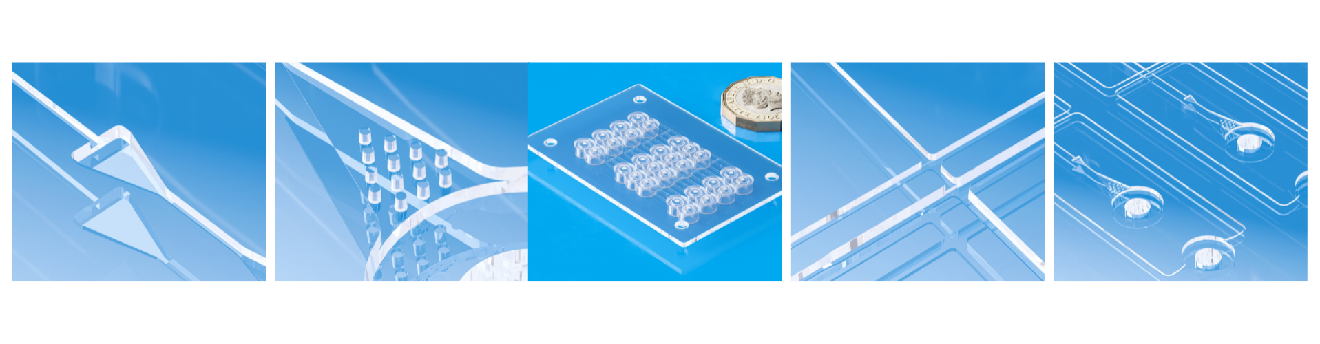
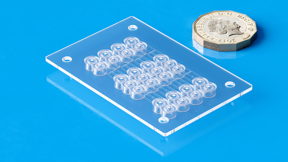
Microfluidics involves the manipulation of fluids at sub-millimeter scales, typically in channels ranging from 10 to 100 micrometers. These platforms, often fabricated from materials like PDMS (polydimethylsiloxane), allow for precise control over fluid movement, enabling functions like mixing, droplet generation, and sorting within a compact lab-on-a-chip system (Sackmann et al., 2014).
The Importance of Single-Cell Analysis
Traditional bulk assays mask cell-to-cell variation, averaging out key biological signals. In contrast, single-cell analysis reveals the heterogeneity within tissues and cell populations, aiding in the discovery of rare cell types, lineage tracing, and disease progression mechanisms (Tanay & Regev, 2017).
How Microfluidics Enables Single-Cell Analysis
High-Throughput Cell Isolation: Microfluidic devices enable the encapsulation of individual cells into microdroplets or wells, each acting as a miniaturised reaction chamber. For example, droplet-based systems like Drop-seq and 10x Genomics Chromium allow simultaneous transcriptomic profiling of thousands of cells (Macosko et al., 2015).
Minimised Reagent Use: Due to the small volumes used, microfluidics dramatically reduces reagent consumption—an advantage for high-cost reagents in omics workflows (Whitesides, 2006).
Integrated Processing and Analysis: Platforms like Auto-ICell integrate cell capture, staining, imaging, and analysis into a single workflow, enabling real-time morphological and apoptotic assessments of individual cells in a low-cost, portable format (Zhou et al., 2023).
Real-Time Monitoring and Dynamic Profiling: Advanced microfluidic platforms now support time-resolved single-cell secretion profiling, enabling researchers to study dynamic processes like cytokine release over time (Chen et al., 2025).
Designing and Manufacturing Microfluidic Devices
Design Considerations: Microfluidic device design requires an interdisciplinary approach, incorporating principles of fluid dynamics, materials science, and biological compatibility. Key design elements include:
- Channel Geometry: Determines flow behaviour, shear stress, and mixing efficiency.
- Valve and Pump Integration: Enables fluid routing and precise control in automated systems.
- Cell Trapping Structures: Microwells, hydrodynamic traps, or droplet encapsulation designs are selected based on target applications (e.g., cell counting vs. culture).
- Computer-aided design (CAD) software, such as AutoCAD or SolidWorks, is commonly used for prototyping, followed by simulation tools like COMSOL Multiphysics to model fluid behaviour at microscale levels (Beebe et al., 2002).
Fabrication Techniques: Common methods include:
- Soft Lithography: The most widely used method, involving photolithography to create a master mold followed by casting PDMS (McDonald et al., 2000).
- Injection Moulding and Hot Embossing: Suitable for large-scale production using thermoplastics such as PMMA or COC. These are preferred for commercial or clinical devices due to their robustness and biocompatibility.
- 3D Printing: Emerging as a flexible, low-cost approach for rapid prototyping of microfluidic devices, especially useful for complex or customised architectures (Waheed et al., 2016).
Material Selection: Choice of material impacts biocompatibility, optical transparency, gas permeability, and ease of surface modification. For example: PDMS is optically transparent, flexible, and gas-permeable, but it can absorb small molecules. Thermoplastics (e.g., PMMA, COC) are more chemically inert and suited for mass production but harder to prototype.
Surface treatments (e.g., oxygen plasma, silanisation) are often used to modify wettability and reduce cell adhesion or protein fouling.
Recent Innovations in Single-cell analysis
Recent advancements in single-cell analysis have significantly expanded the scope and depth of biological discovery. Multi-omics techniques, such as CITE-seq (Cellular Indexing of Transcriptomes and Epitopes by Sequencing), enable simultaneous measurement of mRNA and surface proteins at single-cell resolution, offering a more integrated view of cell phenotype and function (Stoeckius et al., 2017). Other innovations, like SHARE-seq, combine single-cell ATAC-seq and RNA-seq to simultaneously capture chromatin accessibility and gene expression, allowing researchers to dissect regulatory dynamics across cell states (Ma et al., 2020).
Spatial transcriptomics technologies, such as Slide-seq (Rodriques et al., 2019) and DBiT-seq (Liu et al., 2020), now integrate transcriptomic data with spatial context, preserving the native architecture of tissues. These methods use barcoded bead arrays or microfluidic delivery of DNA barcodes to map gene expression patterns across tissue sections at near-cellular resolution, opening new avenues for tumor microenvironment analysis and developmental biology.
Additionally, digital microfluidics has emerged as a key platform for improving single-cell assay scalability and precision. This technique, which manipulates discrete droplets using electric fields, enhances reagent efficiency, reduces contamination, and supports automation, thereby enabling portable and integrated single-cell workflows (Chow et al., 2020).
These cutting-edge tools are driving the next generation of single-cell studies, offering higher throughput, spatial awareness, and multi-layered molecular insights that were previously unattainable.
Applications
Cancer Biology: Microfluidics enables the detection and analysis of circulating tumor cells (CTCs), which are rare but informative about metastatic disease and treatment response.
Immunology: By isolating and profiling immune cells, researchers can investigate immune diversity and dysfunction in diseases such as autoimmunity or cancer.
Stem Cell Research: Microfluidic devices allow precise environmental control and high-throughput screening of stem cell differentiation pathways.
Challenges and Outlook
Despite its promise, microfluidic single-cell analysis still faces challenges:
- Scalability and Standardisation: Lack of uniform fabrication protocols hampers reproducibility.
- Data Overload: The vast amount of data generated requires robust computational tools and storage solutions.
- Integration: Full integration of multi-omics (genomics, proteomics, metabolomics) onto a single chip remains a key research goal.
Manufacturing advancements—especially in scalable, low-cost fabrication—are expected to improve the reproducibility and commercial viability of microfluidic solutions. As fabrication becomes more modular and automated, these devices will transition more readily from research labs to clinical diagnostics.
Microfluidics is enabling a new era in single-cell biology by offering precise, scalable, and high-throughput tools for analysing cellular diversity and function. As device designs evolve and integration with computational methods advances, the next decade will likely see these technologies become routine in both research and clinical settings.
At Micro Systems, our team of experts have decades of experiences working on the most challenging microfluidic device projects, with ISO Class 7 Clearnroom facilities and cutting-edge technologies, we can support you from the design stage all through to mass production of microfluidic device. Contact us today!
References
Beebe, D.J., Mensing, G.A., & Walker, G.M. (2002). Physics and applications of microfluidics in biology. Annual Review of Biomedical Engineering, 4, 261–286.
Chen, Y., et al. (2025). “Time-resolved single-cell secretion analysis via microfluidics.” Lab on a Chip, RSC Publishing. https://pubs.rsc.org/en/Content/ArticleLanding/2025/LC/D4LC00904E
Dutta, A., et al. (2024). “New single-cell analysis tech incorporates advanced fiber optics into microfluidic chips.” Phys.org. https://phys.org/news/2024-04-cell-analysis-tech-incorporates-advanced.html
Macosko, E.Z., et al. (2015). “Highly parallel genome-wide expression profiling of individual cells using nanoliter droplets.” Cell, 161(5), 1202-1214.
McDonald, J.C., et al. (2000). “Fabrication of microfluidic systems in poly(dimethylsiloxane).” Electrophoresis, 21(1), 27–40.
Sackmann, E.K., et al. (2014). “The present and future role of microfluidics in biomedical research.” Nature, 507(7491), 181–189.
Tanay, A., & Regev, A. (2017). “Scaling single-cell genomics from phenomenology to mechanism.” Nature, 541(7637), 331–338.
Waheed, S., et al. (2016). “3D printed microfluidic devices: Enablers and barriers.” Lab on a Chip, 16, 1993–2013.
Whitesides, G.M. (2006). “The origins and the future of microfluidics.” Nature, 442(7101), 368–373.
Zhou, Y., et al. (2023). “Auto-ICell: An integrative droplet microfluidic system for real-time single-cell analysis.” arXiv preprint. https://arxiv.org/abs/2311.02927
Stoeckius, M., Hafemeister, C., Stephenson, W. et al. (2017). Simultaneous epitope and transcriptome measurement in single cells. Nature Methods, 14, 865–868. https://doi.org/10.1038/nmeth.4380
Ma, S., Zhang, B., LaFave, L.M. et al. (2020). Chromatin potential identified by shared single-cell profiling of RNA and chromatin. Cell, 183(4), 1103–1116.e20. https://doi.org/10.1016/j.cell.2020.09.056
Rodriques, S.G., Stickels, R.R., Goeva, A. et al. (2019). Slide-seq: A scalable technology for measuring genome-wide expression at high spatial resolution. Science, 363(6434), 1463–1467. https://doi.org/10.1126/science.aaw1219
Liu, Y., Yang, M., Deng, Y. et al. (2020). High-spatial-resolution multi-omics sequencing via deterministic barcoding in tissue. Cell, 183(6), 1665–1681.e18. https://doi.org/10.1016/j.cell.2020.10.026
Chow, Y.T., Chen, C., Zhang, G. et al. (2020). Digital microfluidics for on-demand single-cell analysis. Lab on a Chip, 20(3), 369–380. https://doi.org/10.1039/C9LC00962H